 |
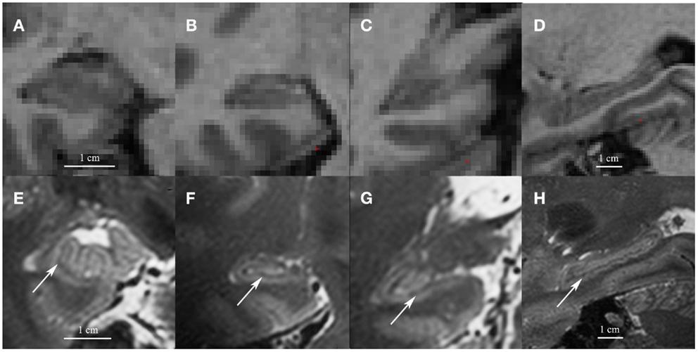
| Coronal images of the head (A), body (B) and tail (C) and a sagittal cross-section of the hippocampus (D) on low resolution 1mm3 T1 1.5 T images (A–D) and on high resolution 0.7 mm3 T2 7 T images (E–H). Note the white matter bands between the dentate gyrus and cornu ammonis on the high resolution T2 images (indicated by arrows). Although we show high resolution 7 T T2 images here, the white matter bands between the dentate gyrus and the cornu ammonis can also be visualized on high resolution T2 3–4 T images |
jueves, 25 de septiembre de 2014
The hippocampus on MRI
Suscribirse a:
Enviar comentarios (Atom)
No hay comentarios:
Publicar un comentario