jueves, 19 de febrero de 2015

domingo, 23 de noviembre de 2014
Diagram of striatal afferent projections from cortex
 |
| Diagram demonstrating the functional organization of A. frontal cortex and B. striatal afferent projections. (A) Schematic illustration of the functional connections linking frontal cortical brain regions. (B) Organization of cortical and subcortical inputs to the striatum. In both (A) and (B), the colors denote functional distinctions. Blue: motor cortex, execution of motor actions; green: premotor cortex, planning of movements; yellow: dorsal and lateral prefrontal cortex, cognitive and executive functions; orange: orbital prefrontal cortex, goal-directed behaviors and motivation; red: medial prefrontal cortex, goal-directed behaviors and emotional processing. (Reproduced with permission from Haber, 2003, fig 3.) |
 |
| Schematic showing anatomy of the striatum (a). Representative lateral (b) and medial (c) illustrations of cortical areas and their connections to the striatum. The colored segment in the striatum represents the area of the striatum receiving projections from the cortical area of the same color. Abbreviations: AC, anterior cingulate cortex; dlPFC, dorsal lateral prefrontal cortex; FEF, frontal eye field; lOFC, lateral orbitofrontal cortex; MC, motor cortex; PPC, posterior parietal cortex; SMA, supplementary motor area; SSC, somatosensory cortex. Compiled from Alexander et al. (1986). (Reproduced from Utter and Basso, 2008, Fig.2) |
domingo, 5 de octubre de 2014
Corpus callosum
The corpus callosum (CC) links the cerebral cortex of the left and right cerebral hemispheres and is the largest fibre pathway in the brain.
Gross anatomy
The corpus callosum is ~10cm in length and is C-shaped, like most of the supratentorial structures, in a gentle upwardly convex arch.
It is divided into four parts (anterior to posterior):
- rostrum (continuous with the lamina terminalis)
- genu
- trunk/body
- splenium
Relations
Immediately above the body of the CC, lies the interhemispheric fissure in which runs the falx cerebri, the anterior cerebral vessels. The superior surface of the CC is covered by a thick layer of grey matter known as the indusium griseum.
On either side, the body is seperated from cingulate gyrus by the callosal sulcus.
Attached to the concave undersurface of the CC is the septum pellucidum anteriorly, and the fornix and its commissure posteriorly.
Fibre tracts
Although the CC can be seen as a single large fibre bundle connecting the two hemispheres, an number of individual fibre tracts can be identified. These include:
- genu: forceps minor : connect medial and lateral surfaces of the frontal lobes
- rostrum: connecting the orbital surfaces of the frontal lobes
- trunk (body): pass through the corona radiata to the surfaces of the hemispheres
- trunk and splenium: tapetum; extends along the lateral surface of the occipital and temporal horns of the lateral ventricle
- splenium: forceps major; connect the occipital lobes
These connections can also be divided into :
- homotopic connections those that link similar regions on each side e.g. visual fields of motor/sensory areas of the trunk
- heterotopic connections: those that link dysimilar areas
Blood supply
The corpus callosum (CC) has a rich blood supply, relatively constant and is uncommonly involved by infarcts. The majority of the CC is supplied by the pericallosal arteries (the small branches and accompanying veins forming the pericallosal moustache) and the posterior pericallosal arteries, branches from the anterior and posterior cerebral respectively. In 80% of patients additional supply comes from the anterior communicating artery, via either subcallosal artery or median callosal artery.
- subcallosal artery (50% of patients) is essentially a large version of a hypothalmic branch, which in addition to supplying part of the hypothalamus also supplies the medial portions of the rostrum and genu
- median callosal artery (30% of patients) can be thought of as a more extended version of the subcallosal artery, in that it travels along the same course, supplies the same structures but additionally reaches the body of the corpus callosum
- posterior pericallosal artery (also known as splenial artery) supplies a variable portion of the splenieum. Its origin is inconstant, arising from P3 or branches there of
jueves, 25 de septiembre de 2014
The hippocampus on MRI
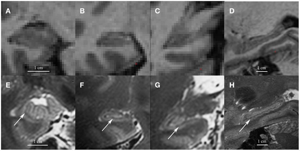 |
| Coronal images of the head (A), body (B) and tail (C) and a sagittal cross-section of the hippocampus (D) on low resolution 1mm3 T1 1.5 T images (A–D) and on high resolution 0.7 mm3 T2 7 T images (E–H). Note the white matter bands between the dentate gyrus and cornu ammonis on the high resolution T2 images (indicated by arrows). Although we show high resolution 7 T T2 images here, the white matter bands between the dentate gyrus and the cornu ammonis can also be visualized on high resolution T2 3–4 T images |
viernes, 18 de abril de 2014
University of Winsconsi's Neuroanatomy Resources
Spinal cord: Global Spinal cord
Spinal cord levels Virtual Brain stem
Thalamus/Hypothalamus/Basal ganglia identifications
Neurosciences video demonstrations Radiology reference page Ventricles Neuroanatomy identifications CNS Development Sleep (ppt) Learn@UW Clinical gallery Coma (ppt)
Week Number Self Learning 2 : Jan 23 - 27 Blood supply
Spinal cord levels
lunes, 31 de marzo de 2014


Suscribirse a:
Entradas (Atom)